
Histology and Microscopy

The Histocore facility provides histology services to university departments, hospital-based research groups, and external partners in the medical and pharmaceutical sectors.
We offer comprehensive expertise in histology, supporting a wide range of research applications, and actively collaborate with industry partners to develop and test new methods, workflows, and therapeutic approaches.
Depending on project needs, we perform a variety of stainings, including immunohistochemistry using the fully automated Bond Rx stainer, as well as immunofluorescence (IF) services.
The histocore facility is a collaboration between BRIC at the University of Copenhagen and the Finsen Laboratory at Rigshospitalet.
For more details, please refer to our workflow, price list, and equipment catalogue.
We recommend contacting the histocore facility at least one month in advance of a project deadline to ensure timely completion.
Step 1: Contact the histocore facility by e-mail (external users) or PPMS (internal BRIC users)
Step 2: Meet with one of our core facility scientists to plan and align on your project
Step 3: Complete a request sheet outlining the agreed project services
Step 4: Your projected is completed according to your specifications
The total expected processing time of steps 1-4 is one month. Longer processing time during peak periods, as main holiday periods may apply.
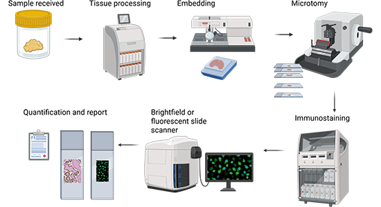
Tissue Collection
The process begins with the collection of tissue samples, typically preserved in a fixative solution within a container. This step ensures the preservation of tissue morphology and prevents degradation.
Tissue Processing
The fixed tissue is then processed using an automated tissue processor. This machine dehydrates the tissue by passing it through a series of alcohols and clears it with a solvent e.g. tissue clear, preparing the tissue for embedding.
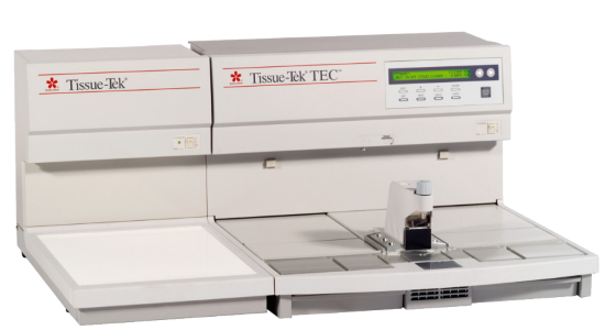
Tissue Embedding
After processing, the tissue is embedded in paraffin wax using an embedding station. This step solidifies the tissue into a block, which can easily be sectioned for microscopic examination.
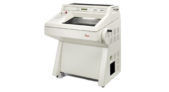
Microtomy
The paraffin embedded tissue block is then sectioned using a microtome. Thin slices of the tissue, usually 3-5 microns, are cut and placed on microscope slides for staining.
Staining
Tissue slides are stained using, for example, an automated staining machine for Hematoxylin and Eosin (H&E) to highlight various tissue components.
Depending on the analysis requirements, additional stains can be applied, including immunohistochemical stains, using the fully automated Bond Rx stainer.
For simultaneous detection of multiple proteins, fluorescence Multiplex is required, such as assays from SignalStar™, which allows fully automated analysis up to 8-plex.
Additionally, RNAscope can be used if the project aims to examine RNA levels, with the possibility of co-detecting both RNA and protein.
Imagining, Quantification and Reporting
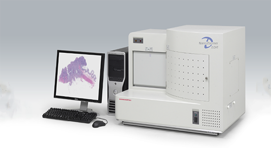
After staining, the slides are analyzed using a microscope, often equipped with imaging capabilities. We use the digital slide scanner Nanozoomer, for whole slide imaging.
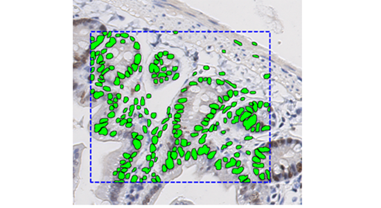
The final images and data from the stained sections are analyzed and interpreted using Visiopharm software, specifically Visiopharm’s AI-powered image analysis platform.
The results are documented in a report, which is used for diagnostic or research purposes.
Service based
The BondRx can be used for complete IHC, RNAScope, FISH, multiplexing and other tests.
For immunohistochemical stainings you need to consider the following:
- Pretreatment
- Antibody concentration
- Secondary antibody
- Detection system
Manufacturer: Leica

The final images and data from the stained sections are analyzed and interpreted using Visiopharm software, specifically Visiopharm’s AI-powered image analysis platform. The results are documented in a report, which is used for diagnostic or research purposes.
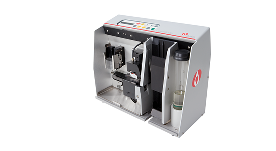
Automated HE stainer ensures repeatable HE stainings of high quality.
Manufacturer: Epredia
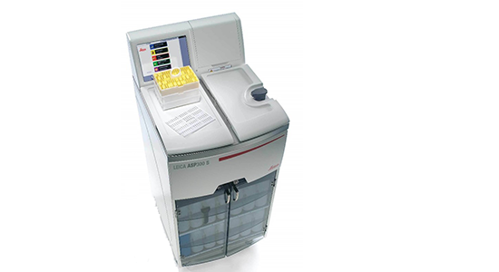
For tissue processing.
The tissue must be fixated in PFA in 24 hours, thereafter kept in 70 % ethanol. Bring samples to cupboard in room 3.3.42.
Manufacturer: Leica
Access: Service based
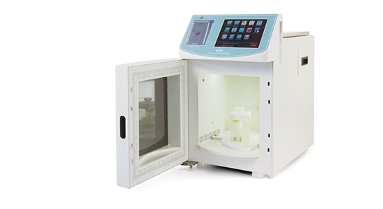
For Antigen retrieval, defalcification.
Manufacturer: Milestone
Access: Service based
Manufacturer: Dako
Access: Service based
Access based
The following instruments are access-based instruments, which can be booked and used after thorough training from histocore personnel.
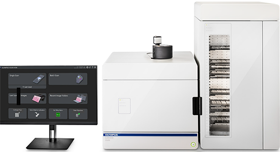
The SLIDEVIEW VS200 system offers high-resolution and high-throughput slide scanning for advanced research applications, such as neuroscience, cancer and stem cell research, and spatial biology.
With the flexibility of five imaging modes, multiplexing capabilities, and multiple magnifications, the system delivers outstanding image quality for quantitative analysis.
Manufacturer: Olympus
Manufacturer: Hamamatsu
Model: HM 355S Automatic Microtome
Manufacturer: Thermo scientific
Model: HM 355S Automatic Microtome
Manufacturer: Microm
Model: RM2255 Fully Automated Rotary Microtome
Manufacturer: Leica
Model: SM2000 R Sliding microtome
Manufacturer: Leica

Manufacturer: Leica
Manufacturer: Sakura
For incubating slides, before staining.
Manufacturer: Buch & Holm
Prices
| Institute | Price |
| UCPH internal | 500 DKK |
| Industry | 1400 DKK |
| Min cost | 15 min |
| Visiopharm software | Price |
| Access based use | Write email for estimated price |
| Service based use | Write email for estimated price |
| Consumables | Price |
| HE staining | 125 DKK pr basket |
| Microtomeblades | 30 DKK pr unit |
| Slide | 2,5 DKK pr unit |
|
IHC/IF/RNAScope |
Write email for estimated price |
NanoZoomer-XR Digital slide scanner C12000-01 – Hamamatsu
| Institute | Price pr hour |
| UCPH internal 9-16 | 150 DKK |
| UCPH internal 16-9 | 100 DKK |
| Industry all day | 200 DKK |
SLIDEVIEW VS200 Universal Whole Slide Imaging Scanner – Olympus
| Institute | Price pr hour |
| UCPH internal | 320 DKK |
| External academia | 640 DKK |
| Industry | 1300 DKK |
| Institute | Price pr hour |
| External/Industry | 400 DKK |
Contact
Histology and Microscopy
Mail: histocore@finsenlab.dk
Phone: +45 9356 5859
BRIC - Biotech Research & Innovation
Centre Ole Maaløes Vej 5
DK-2200 Copenhagen
Room 3-3-4 (Lab) or
3.3.25 (Office).

Mia Kristine Høg
Lead Core Facility Scientist
Research Biomedical laboratory Scientist
Mail: histocore@finsenlab.dk

Manja Nordberg
Core Facility Scientist
Mail: histocore@finsenlab.dk

Lars H. Engelholm
Head of Facility, Group Leader
Mail: lhe@finsenlab.dk