Kristensen Group
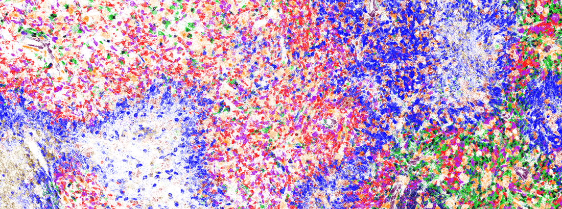
The Kristensen Group is located at the Bartholin Institute, which is part of Department of Pathology at Rigshospitalet, Copenhagen University Hospital.
Glioblastoma is the most frequent and malignant type of brain cancer. Patients diagnosed with glioblastomas are among those cancer patients that have the poorest overall survival. By understanding and overcoming the mechanisms of aggressiveness and resistance in glioblastoma, we strive to discover novel molecular targets for more effective therapies for the patients.
The glioblastoma microenvironment is highly heterogenous, resulting in diverse mechanisms of aggressiveness and resistance across different regions of the tumor. Adding to the complexity, there is a high frequency of non-tumor cells in glioblastomas, which leads to pro-tumorigenic crosstalk between tumor cells and non-tumor cells. With this in mind, we work to understand the underlying drivers of aggressiveness and resistance.
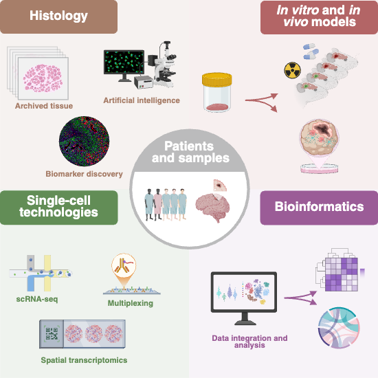
Our research utilizes patient tissue cohorts, single-cell technologies and patient-derived in vitro and in vivo preclinical models to generate translational insights, with the ultimate goal of identifying novel therapeutic strategies for glioblastoma patients.
Spatial insight into the microenvironment
- Infiltrating glioblastoma cells are expressing Notch and synaptic genes, as identified using single cell spatially resolved transcriptomics. Harwood et al., Nature Communications (2024), PMID: 39251578 DOI: 1038/s41467-024-52167-y
- Multiomic profiling of glioblastoma metabolic lesions reveals complex intratumoral genomic evolution and dipeptidase-1-driven vascular proliferation. Anand et al, Neuro Oncology (2025), PMID: 40319462 DOI: 10.1093/neuonc/noaf071
- Checkpoint markers are differentially expressed in the normoxic and hypoxic microenvironment. Petterson et al, Brain Pathology (2023), PMID: 36093941 DOI: 1111/bpa.13111
- CD204+ TAMs accumulate in perivascular and perinecrotic niches and correlate with an interleukin-6 enriched inflammatory profile. Sørensen et al, NAN (2022), PMID: 34713474 DOI: 1111/nan.12772
Microenvironment, tumor recurrence and prognosis
- Surgery - in a patient-derived glioblastoma resection model - promotes early recurrence, leads to recurrences that harbor a large number of tumor-associated microglia and -macrophages, and gains a more aggressive phenotype than primary glioblastomas. Knudsen et al, Neuro Oncology (2022), PMID: 34964899 DOI: 1093/neuonc/noab302
- Microglia induce an interferon-stimulated gene expression profile in glioblastoma and increase glioblastoma resistance to chemotherapy, Sørensen et al, NAN (2024) PMID 39558550 DOI: 1111/nan.13016
- Poor prognosis is observed in patients receiving chemotherapy, when large number of CD204+ TAMs are present in the primary tumor. Sørensen et al, NAN (2018) PMID: 28767130 DOI: 1111/nan.12428
- GDNF/GFRA1 signaling contributes to chemo- and radioresistance in glioblastoma, Avenel et al, Sci Rep (2025), PMID: 39085346 DOI: 1038/s41598-024-68626-x
Mutational profile influences the microenvironment
- D-2-HG in IDH-mutant astrocytic gliomas suppresses the host immune system. Zhang et al, Clin Cancer Research (2018), PMID: 30006485 DOI: 10.1158/1078-0432.CCR-17-3855
- IDH mutation is associated with diminished levels of TIM-3+ cells and fewer interactions between TIM-3+ T cells and galectin-9+ TAMs, suggesting reduced activity of the galectin-9/TIM-3 immune checkpoint pathway in IDH-mutant astrocytic gliomas. Sørensen et al, Brain Pathology (2020), PMID: 33244787 DOI: 10.1111/bpa.12921
Identification of novel therapeutic strategies for brain cancer
Glioblastomas are characterized by extensive areas with hypoxia, peri-necrotic palisades and microvascular proliferation, which are hallmarks of glioblastoma. In the outer part of glioblastomas, highly infiltrative tumor cells are located, that infiltrate the brain and interact with cells of the brain parenchyma.
These infiltrating tumor cells are often left after surgical resection and lead to tumor recurrence. In this project we work to understand the spatial biology of glioblastoma and how it changes during development of the disease – from the core of the tumors to the infiltrative outer part of the tumors and to recurrence. An important part of this is to understand cellular crosstalk and its impact on aggressiveness and resistance, e.g. the impact of tumor-associated microglia and macrophages on tumors.
We use single cell technologies with an emphasis on spatial profiling and high-end immunohistochemical multiplexing. Our goal is to identify potential novel targets we can test in our pre-clinical models.
Therapy resistance in glioblastoma: tumor cells and the surrounding microenvironment
We interrogate the existence, characteristics and role of different clinically important subtypes of tumor associated microglia and macrophages in glioblastoma.
Methodologically we use e.g. single cell technologies to investigate fresh patient glioblastoma tissue combined with genetically engineered mice models to identify and functionally investigate cellular crosstalk and resistance mechanisms.
Our goal is to identify potential novel therapeutic strategies to prevent tumor promoting cellular crosstalk.
Improved pre-clinical models
Novel therapies are rarely investigated in the context of tumor resection in preclinical models - although chemo-radiation therapy is given after resection in patients. However, early recurrences of glioblastoma contain a high amount of inflammatory and reactive cells, which interact with and affect tumor cells.
To model these early timepoints of recurrence, we work with 3D in vitro models and pre-clinical models with tumor resection to interrogate the tumor cell phenotype and the microenvironment.
Our goal is to identify novel targets that can eventually lead to prevention of recurrence of glioblastoma.
We use brain tumor tissue from patients for in vitro (e.g., cell, spheroid or organoid cultures) and in vivo (primarily mouse) pre-clinical models, confocal time-lapse microscopy, flow cytometry as well as single cell/nuclei sequencing.
To discover novel targets, biomarkers and resistance mechanisms, we use patient cohorts, spatial profiling, immunohistochemistry, computer-based biomarker analysis, NGS, genome wide methylation profiling, mRNA profiling and proteomics.
Core facilities
To obtain a better understanding of how cells and tissue compartments are spatially organised in brain tumor tissue, we utilize immunofluorescence multiplexing and deep spatial profiling.
Equipment for these techniques are available as core facilities, with open access, and are located at the Bartholin Institute.
- Vectra Polaris
- Automated Quantitative Pathology Imaging System
- Thunder Imager - Advanced 3D cell culture assays
- Zeiss LSM 900 confocal microscope
- Laser dissection microscopy
- Visiopharm imaging software – image analysis powered by AI Deep Learning
- The Nanostring GeoMx platform for Digital Spatial Profiling.
Selected publications
Multiomic profiling of glioblastoma metabolic lesions reveals complex intratumoral genomic evolution and dipeptidase-1-driven vascular proliferation. Anand A, Petersen JK, Andersen LVB, Burton M, Oudenaarden CRL, Larsen MJ, Erichsen PA, Pedersen CB, Poulsen FR, Grupe P, Kruse TA, Thomassen M, Kristensen BW. Neuro Oncol. 2025 May 4:noaf071. doi: 10.1093/neuonc/noaf071. Online ahead of print. PMID: 40319462.
Glioblastoma cells increase expression of notch signaling and synaptic genes within infiltrated brain tissue. Harwood DSL, Pedersen V, Bager NS, Schmidt AY, Stannius TO, Areškevičiūtė A, Josefsen K, Nørøxe DS, Scheie D, Rostalski H, Lü MJS, Locallo A, Lassen U, Bagger FO, Weischenfeldt J, Heiland DH, Vitting-Seerup K, Michaelsen SR, Kristensen BW. Nat Commun. 2024 Sep 9;15(1):7857. doi: 10.1038/s41467-024-52167-y. PMID: 39251578.
Avenel ICN, Ewald JD, Ariey-Bonnet J, Kristensen IH, Petterson SA, Thesbjerg MN, Burton M, Thomassen M, Wennerberg K, Michaelsen SR, Kristensen BW. GDNF/GFRA1 signaling contributes to chemo- and radioresistance in glioblastoma. Sci Rep. 2024 Jul 31;14(1):17639. doi: 10.1038/s41598-024-68626-x. PMID: 39085346; PMCID: PMC11292001.
SA Petterson, MD Sørensen, M Burton, M Thomassen, TA Kruse, SR Michaelsen, BW Kristensen. Differential expression of checkpoint markers in the normoxic and hypoxic microenvironment of glioblastomas, Brain Pathology, 2023 Jan; 33 (1):e13111, PMID: 36093941
AM Knudsen, B Halle, O Cédile, M Burton, C Baun, H Thisgaard, A Anand, C Hubert, M Thomassen, SR Michaelsen, BB Olsen, RH Dahlrot, R Bjerkvig, JD Lathia, BW Kristensen. Surgical resection of glioblastomas induces pleiotrophin-mediated self-renewal of glioblastoma stem cells in recurrent tumors. Neuro Oncol. 2022 July 1;24(7):1074-1087, PMID: 34964899
MD Sørensen, BW Kristensen. Tumour-associated CD204+ microglia/macrophages accumulate in perivascular and perinecrotic niches and correlate with an interleukin-6 enriched imflammatory profile in glioblastoma. Neuropathol Appl Neurobiol. 2022 Feb;48(2):e12772, PMID: 34713474
M.D. Sørensen, R.H. Dahlrot, H.B. Boldt, S. Hansen and B.W. Kristensen. Tumor-associated microglia/macrophages predict poor prognosis in high-grade gliomas and correlate with an aggressive tumor subtype. Neuropathol Appl Neurobiol, Feb;44(2), 185-206, 2018. PMID 28767130.

We host the SNOG - NORTHERN LIGHTS NEUROSCIENCE SYMPOSIUM taking place 9-10 April 2026 in Copenhagen with Professor Bjarne Winther Kristensen as Congress President.
27 November 2024
Researchers can now identify cancer cells leading to tumor recurrence
September 2022
Professor Bjarne Winther Kristensen receives grant from NEYE-Foundation for the project: Targeting glioblastoma recurrence by novel insight into the post-surgical microenvironment in patients (Danish).
2020
Kristensen group hosts the 12th European Congress of Neuropathology with Professor Bjarne Winther Kristensen as Congress President.
15 November 2016
Danske forskere finder roden til hjernekræftens onde (Danish).
Danish Cancer Society
Novo Nordisk Foundation
Danish Council for Independent Research
Lundbeck Fonden
Beckett Fonden
Dansk Kræftforskningsfond
Neye
Toyota Fonden
Harboefonden
